Sonoscan
Gen-6
The Sonoscan Gen-6 is a C-mode scanning acoustic microscope. The Sonoscan Gen-6 is capable of scanning a 300mm wafer. The Sonoscan Gen-6 includes PolyGate technology with Multi-Gate and Probing-Gate functions.[1][2]
Plate 01Gen6 Setup Part1of2
Nordson SonoscanWatch on YouTubePlate 02Gen6 Setup Part2of2
Nordson SonoscanWatch on YouTube
Wafer size
300mm
Optics
Open-access illuminated scanning area[1]
How it works
The Sonoscan Gen-6 uses PolyGate technology with Multi-Gate and Probing-Gate functions for single and multi-focus imaging. The instrument supports up to one hundred gates per channel and uses a two gigasamples-per-second sampling rate.[1][2]
The Sonoscan Gen-6 uses an inertially balanced linear motor scanner, a tower-mounted scan reference platform, and a sample fixture. The instrument also supports water recirculation and optional inline temperature control.[1][2]
Where it fits in the process flow
Applications
The Sonoscan Gen-6 supports inspection of stacked die parts through SonoSimulator and supports digital image analysis for quantitative accept or reject criteria. The instrument also supports virtual rescanning for complete analysis after the sample is no longer available.[1][2]
- Nondestructive failure analysis
- Process development
- High-Rel qualification for a military application
- Low/medium volume screening
- Microelectronics
- MEMS
- SSL LEDs
- Power modules
- Solar
- HighTech materials
What do the numbers mean?
Wafer handling2
Optics & imaging1
Control & software5
- Software analysis tools
- SonoSimulator, Digital Image Analysis (DIA), and Virtual Rescanning Mode (VRM)[1]
Configuration & options11
- Technology
- PolyGate™ technology with Multi-Gate™ and Probing-Gate™ functions capable of single and multi-focus imaging[1]
- Data analysis
- Digital Image Analysis (DIA) quantifies acoustic data and supports automatic accept/reject criteria[1]
- Rescanning
- Virtual Rescanning Mode (VRM) stores comprehensive data for complete analysis after the sample is no longer available[1]
Where are the manuals?
Generated from public-source data on file. Enter your email to access — nothing is published; details are routed privately.
Not publicly documented
Field notes
No research found yet — worked with this tool? Share what you know.
Frequently asked questions
Does the sample need any special preparation before inspection?General reference — not yet source-verified
No special preparation is typically required, but the sample must be compatible with the coupling fluid (usually deionized water). The surface should be clean and able to be wetted by the fluid. Some samples may need to be dried after inspection.
How does acoustic microscopy differ from X-ray inspection?General reference — not yet source-verified
Acoustic microscopy is sensitive to mechanical discontinuities (gaps, cracks) while X-ray detects density variations and absorbs through materials. Both are complementary: X-ray sees metal structures and voids, while acoustic microscopy is better for finding delamination and cracks in polymer and adhesive layers.
Is the inspection method destructive to the sample?General reference — not yet source-verified
No, scanning acoustic microscopy is a non-destructive inspection method. The sample is immersed in a fluid couplant but experiences no damage, so tested parts can be returned to service or used for further analysis.
Not publicly documented
The following facts about the Gen-6 are absent from this record as of this revision. First-hand knowledge or a citation closes a gap; every submission is reviewed before publication.
No publicly documented production dates or lifecycle milestones (introduction, end of production, EOL) for the Gen-6 are on record.
Answerable by: OEM historical records or a trade-press announcement
No publicly documented variants, configuration options, or revision breakpoints of the Gen-6 are on record.
Answerable by: an OEM product catalog or an engineer who ordered or specified the tool
The control-system platform and OS era of the Gen-6 are not on record.
Answerable by: an engineer who operated it or OEM installation records
No publicly documented failure modes or field errata for the Gen-6 are on record.
Answerable by: a field service engineer, process engineer, or maintenance technician
The process node or technology generation of the Gen-6 is not on record.
Answerable by: an OEM datasheet or a fab qualification report
No research found yet — worked with this tool? Share what you know.
Sources & citations
Sources (3)Every fact above is drawn from these public sources
- [1]ncf.uic.edu — ncf.uic.eduncf.uic.edu
- [2]https://ncf.uic.edu/wp-content/uploads/sites/819/2022/04/Scanning-Acoustic-Microscope-Manual.pdf — ncf.uic.eduncf.uic.edu
- [3]Instruments and Services | Nanotechnology Core Facility | University of Illinois Chicago — ncf.uic.eduncf.uic.edu
Last updated Oct 3, 2026.
Compare with similar tools
View all →The Rudolph E25 System is an automated wafer edge top and edge normal inspection system for 300 mm wafers.
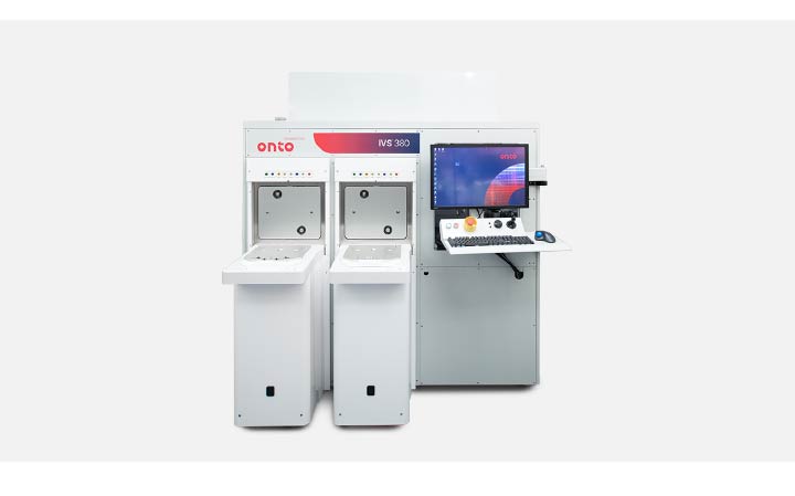
The IVS 380 System performs overlay, CD, and height metrology for 300mm wafers.

The KLA Surfscan SP 2 is a particle defect inspection system for semiconductor wafers.
The KLA .204 PSL Wafer is an 8-inch NIST-traceable reference wafer compatible with Surfscan 6xy0 and Sp1 particle counters.